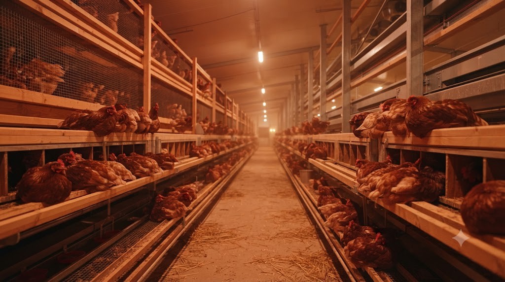
Egg quality is the supreme metric of commercial viability and consumer trust in table egg production. In competitive agricultural markets, every harvested egg is evaluated against rigorous commercial grading parameters codified by international agricultural standards (such as USDA Grade AA, Grade A, and Grade B, or European Union Class A standards). Down-grading or discarding eggs due to exterior shell imperfections, microbial penetration, or internal inclusions imposes substantial financial losses on poultry producers, eroding operating profit margins by 5% to 15% annually.
Diagnosing egg defects requires deep veterinary pathology comprehension. An egg is not a static manufactured commodity; it is a sensitive biological readout of the hen’s instantaneous health, nutritional status, endocrine balance, and environmental stress. This comprehensive clinical guide examines the physiological etiology, diagnostic indicators, and actionable preventative protocols for both external shell defects and internal albumen and yolk anomalies.
🥚
Shell Pathology
External Eggshell Defects: Biophysical Etiology and Prevention
External defects compromise the egg’s primary physical protective armor. Because the shell gland deposits roughly 2.2 grams of calcium carbonate within an intense 16-to-18-hour calcification window, any physiological disturbance during this period manifests as distinct structural deformities.
1. Soft-Shelled and Completely Shell-Less Eggs
Soft-shelled eggs possess only a paper-thin calcified glaze over the shell membranes, while completely shell-less eggs consist solely of albumen and yolk encased in flexible fibrous membranes. Etiological factors include:
- Severe Nutritional Deficits: Inadequate dietary calcium, severe phosphorus imbalance, or deficiency in active Vitamin D3 ($1,25(OH)_2D_3$), which prevents calcium absorption across enterocytes.
- Premature Oviposition Triggered by Panic: Sudden loud noises, predator incursions, or handling during late evening induce an acute adrenaline surge. Adrenaline triggers immediate, violent uterine smooth muscle contractions that expel the developing egg hours before calcification is complete.
- Viral Pathology (Egg Drop Syndrome – EDS’76): Caused by an avian adenovirus, EDS’76 attacks the uterine mucosal plicae, causing severe localized inflammation and completely shutting down calcium secretion without producing overt systemic illness in the bird.
2. Body-Checked (Repaired Fissure) Shells
A body-checked egg displays a raised, irregular ridge or corrugated band running equatorially around the shell. A body check occurs when an eggshell is cracked mechanically while inside the hen’s uterus during early calcification (typically 10 to 14 hours after ovulation). The cracked fragments remain held in place by the shell membranes, and the uterine shell gland subsequently deposits a secondary layer of calcite over the crack, sealing the fracture line before oviposition. The primary cause is physical collision: overcrowding on perches, inadequate roosting space, or flight panics during late afternoon.
3. Corrugated, Wrinkled, and Misshapen Shells
Wrinkled or corrugated shells feature prominent longitudinal or circular folds across the surface. This defect is a hallmark clinical sign of Infectious Bronchitis Virus (IBV). IBV possesses a specific tropism for the ciliated and secretory epithelial cells of the oviduct. Even after the flock recovers from mild respiratory symptoms, IBV causes chronic, permanent atrophy and scarring of the magnum and uterine mucosal folds. Because the damaged oviduct cannot properly regulate albumen plumping pressure, the shell membranes wrinkle, and subsequent calcification permanently casts the wrinkled deformity in stone.
| Egg Defect Type | Visual Appearance | Primary Anatomical Origin | Underlying Biological Cause | Recommended Corrective Action |
|---|---|---|---|---|
| Soft-Shelled / Shell-Less | Flexible membrane; little/no calcite | Uterine Shell Gland | Severe Ca/D3 deficit; EDS’76 virus; stress expulsion | Add coarse limestone; check D3 levels; review flock biosecurity |
| Body-Checked Shell | Raised equatorial seam / ridge | Uterus (early calcification) | Mechanical trauma; perching collision in late evening | Increase perching space; eliminate evening disturbances |
| Corrugated / Wrinkled | Wavy, creased surface contours | Magnum & Uterus | Infectious Bronchitis Virus (IBV) damage | Vaccinate with homologous IBV strains during pullet rearing |
| Pimpled / Calcified Rough | Granular sandpaper calcium bumps | Uterine Lumen | Excess dietary calcium; foreign dust; aged hens | Balance Ca:P ratio; clean feeder lines; check limestone size |
| Blood Spots | Bright red dot on yolk surface | Ovarian Stigma | Follicular capillary rupture during ovulation | Stabilize lighting; check Vitamin K3; minimize flock panic |
| Meat Spots | Brown/tan speck in dense albumen | Magnum Epithelium | Sloughed oviduct tissue; oxidized blood clot | Review genetic strain selection; minimize physiological stress |
| Watery Albumen | Runny, flat white; low Haugh unit | Magnum Glandular Folds | Aged hens; elevated coop storage temps; IBV infection | Harvest eggs twice daily; store at 10-12°C; ensure dietary protein |
🥚
Internal Defects
Internal Egg Inclusions: Blood Spots, Meat Spots, and Albumen Breakdown
While consumers cannot see internal inclusions through the shell at retail purchase, finding internal defects destroys consumer confidence and triggers regulatory grade penalties.
Blood Spots: The Ovarian Capillary Rupture
A blood spot appears as a distinct droplet of bright red blood affixed directly to the surface of the yolk or floating in the perivitelline space. Blood spots do not indicate a fertilized embryo; rather, they are caused by localized microvascular hemorrhage during ovulation.
Normally, the ovarian follicle ruptures cleanly along the avascular stigma line. However, if capillary networks cross the stigma or if the hen experiences high blood pressure or acute fright during the LH surge, a capillary ruptures, spraying a droplet of blood onto the yolk just as it drops into the infundibulum. Deficiencies in Vitamin K3 (menadione)—which is required for hepatic synthesis of prothrombin and blood-clotting factors—greatly exacerbate blood spot incidence. Furthermore, brown-shelled hybrid strains naturally exhibit a higher genetic predisposition to blood spots (typically 1.5% to 3.0%) compared to white Leghorns (under 0.5%).
Meat Spots: Tissue Desquamation and Oxidation
Meat spots are small, solid flecks of brownish, tan, or greyish tissue suspended within the thick albumen. Historically believed to be bits of sloughed oviductal mucosa, advanced histological assays reveal that the vast majority of meat spots are actually aged blood spots that underwent chemical oxidation. When an ovarian blood droplet is exposed to the acidic pH of the magnum and the oxidative action of oviductal secretions, hemoglobin iron oxidizes from bright red ferro-hemoglobin into brown ferri-hemoglobin, resulting in a dark meat spot.
Watery Albumen and The Haugh Unit Metric
Interior egg quality is quantified scientifically by measuring the Haugh Unit (HU), established by Raymond Haugh in 1937. The Haugh unit mathematical formula correlates the height of the thick albumen (measured in millimeters with an electronic micrometer) with the mass of the egg (grams):
Haugh Unit = 100 * log10 [ h – 1.7 * w^0.37 + 7.6 ]
An elite fresh egg scores above 72 Haugh Units (USDA Grade AA). As eggs age, or if hens suffer from chronic respiratory infections or excessive dietary vanadium, thick albumen breaks down through lysozyme-ovomucin complex dissociation, causing the albumen to spread flat and watery. Storing eggs at 50°F to 55°F (10°C to 12.8°C) with 75% relative humidity immediately arrests albumen degradation.
🥚
Grading Technology
Industrial Candling and Crack Detection Technologies
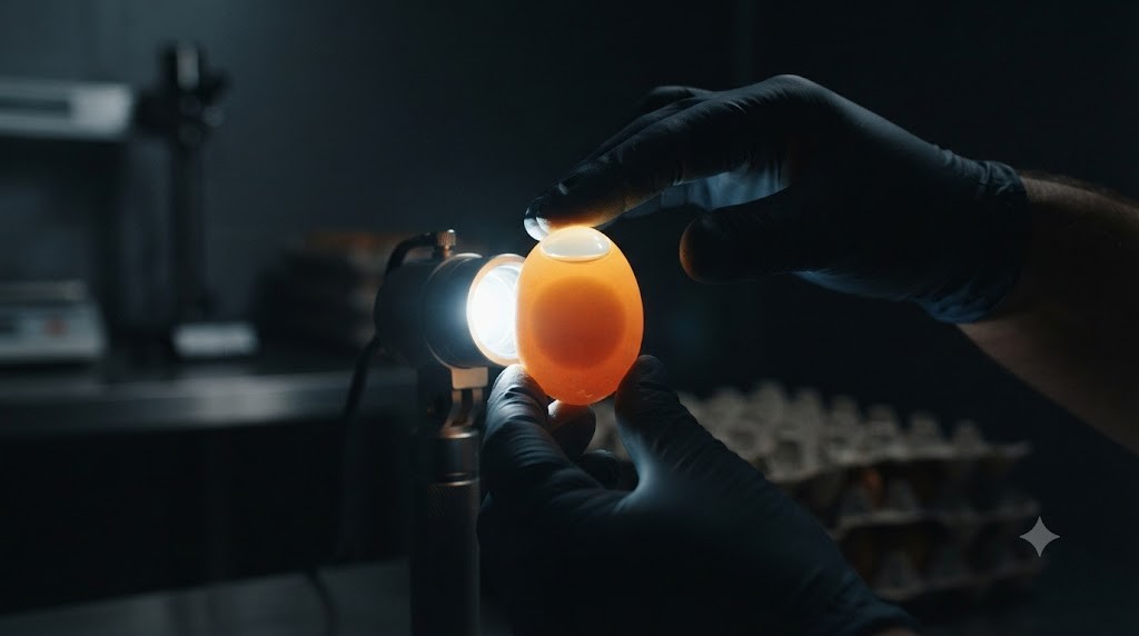
Modern egg grading facilities process hundreds of thousands of eggs per hour utilizing sophisticated automated optical and acoustic inspection arrays:
High-Speed Optical Candling
Eggs travel over high-intensity LED light beams while rotating along their long axis. Automated high-resolution machine-vision cameras capture digital images of each egg, instantaneously detecting internal air cell diameter, yolk shadow mobility, blood spots, and internal inclusions, automatically ejecting defective eggs onto diverted packaging lanes.
Acoustic Resonance Crack Detection
Micro-cracks in the shell are often invisible to the naked eye. Automated grading machines deploy delicate acoustic tap probes that strike the eggshell at 16 to 24 locations in fractions of a second. High-frequency acoustic sensors analyze the resonant acoustic frequency of the vibration: an intact crystalline calcite shell vibrates at a clear, high pitch, whereas a cracked or body-checked shell produces a muffled, distorted harmonic frequency, triggering immediate robotic pneumatic rejection.
By implementing proactive flock vaccination, precision mineral nutrition, stress-free housing protocols, and state-of-the-art optical candling, commercial poultry operations successfully minimize egg defects, protect consumer loyalty, and capture maximum market value across every crate of eggs produced.
🥚
Cuticle Science
Cuticle Quality Staining and Microbial Resistance: The MST Assay
The outermost defense of the newly laid egg is the cuticle—a delicate, nanometer-thin organic layer composed of glycoproteins, polysaccharides, and lipids deposited onto the calcified palisade layer just minutes before oviposition. The cuticle seals the microscopic respiratory pores (which number between 7,000 and 17,000 per egg), preventing opportunistic environmental bacteria (such as Salmonella enterica and Pseudomonas) from penetrating the liquid egg interior while permitting the passive diffusion of respiratory gases.
Quantifying Cuticle Integrity via MST Dye Staining
In quality control laboratories and breeding research, cuticle coverage is evaluated using the standardized MST (Micro-Spectrophotometry) staining assay. Eggs are submerged in a specialized dye solution containing Coomassie Brilliant Blue and Lissamine Green for 60 seconds, then rinsed in deionized water. Because the dye binds selectively to the proteins of the organic cuticle and cannot bind to inorganic calcium carbonate, eggs with complete, robust cuticles stain a deep, uniform emerald green, whereas eggs with patchy or absent cuticles remain chalky white.
Research reveals that cuticle thickness naturally diminishes as hens age, and is severely eroded by aggressive commercial egg washing protocols that use high-pH alkaline detergents and abrasive mechanical nylon brushes. Preserving cuticle integrity through gentle washing and dietary organic mineral supplementation is vital for preventing post-harvest bacterial contamination.
🥚
Yolk Quality
Yolk Coloration Standards and DSM YolkFan Calibration
While yolk color does not directly alter the basic protein content of an egg, it is the primary aesthetic quality trait driving consumer purchase preferences globally. Yolk color is governed entirely by dietary carotenoids; chickens cannot synthesize carotenoids de novo and must absorb them from their feed.
The Biochemistry of Yellow and Red Carotenoids
Yolk color is evaluated commercially using the standardized 15-blade DSM YolkFan (formerly Roche Yolk Color Fan), scoring from pale straw yellow (score 1) to deep reddish-orange (score 15). Achieving deep, vibrant yolk color requires a balanced synergy of two carotenoid classes:
- Yellow Carotenoids (Lutein and Zeaxanthin): Sourced from yellow corn, corn gluten meal, and marigold flower extract (Tagetes erecta). Yellow carotenoids establish the golden baseline color (scores 1 through 7).
- Red Carotenoids (Capsanthin and Canthaxanthin): Sourced naturally from red chili peppers (Capsicum annuum) or nature-identical canthaxanthin. Red carotenoids add warm red saturation, elevating yolk scores from 8 to 14+.
Consumer preferences vary widely across international markets: consumers in Germany, Japan, and Latin America demand deep orange-red yolks (scores 12 to 14), while North American and UK consumers traditionally favor golden-yellow yolks (scores 8 to 10). Nutritionists adjust carotenoid inclusion matrices to target specific retail contracts.
🥚
Microbiology & Food Safety
Microbial Spoilage Pathways: Bacterial Penetration and Rotten Eggs
An intact, freshly laid egg possesses formidable natural antibacterial defenses: the hydrophobic cuticle barrier, the physical shell membranes, alkaline albumen pH (rising from 7.6 to 9.2 within days of lay), and potent antibacterial enzymes including lysozyme, ovotransferrin, and avidin. However, under suboptimal harvesting or washing conditions, opportunistic bacteria can penetrate these defenses, causing severe microbial spoilage.
The Physics of Thermal Siphoning during Egg Washing
The single greatest cause of bacterial contamination in commercial egg packing plants is improper wash water temperature. When an egg is laid, its internal contents are at the hen’s body temperature (approximately 105°F / 40.5°C). As the egg cools, the internal liquid contracts, creating a slight negative pressure that draws ambient air inward through the shell pores, forming the air cell at the blunt end.
If eggs are washed in water that is colder than the internal temperature of the egg, this negative pressure thermal siphon accelerates dramatically: cold wash water containing surface dirt and bacterial pathogens (such as Pseudomonas fluorescens and Salmonella) is sucked directly through the shell pores into the interior albumen. USDA standards mandate that wash water must be at least 20°F (11°C) warmer than the warmest egg (minimum 90°F / 32.2°C) to maintain positive internal pressure, ensuring that fluids are pushed outward through pores rather than sucked inward.
Diagnostic Classification of Bacterial Rots
- Green Rots (Pseudomonas fluorescens): Characterized by luminous, fluorescent green albumen under ultraviolet candling, accompanied by a distinctive sour, fruit-like odor.
- Black Rots (Proteus and Aerobacter): The interior breaks down into a liquefied black mass emitting pungent hydrogen sulfide ($H_2S$) rotten-egg gas. Internal gas expansion frequently causes the egg to explode violently during handling.
- Red and Pin-Spot Rots (Serratia marcescens): Manifest as rusty red rust spots along the vitelline membrane, contaminating food preparations.